HSCA Published X-rays
This article is divided into the following sections:
Authenticity Problems
The first order of business is the authentication of the X-rays. And that authentication is extremely problematic.
Dr. David Mantik, M.D., Ph.D., addresses this problem extensively in his work. Dr. Mantik is both a physicist and medical doctor by education and training, and an oncology radiologist by trade--meaning he studies X-rays for a living, specifically related to the diagnosis and treatment of cancer. He received permission from the Kennedy family to view the X-rays first-hand in the Archives. He did this with his own eyes as well as the aid of an optical densitometry machine, which measures the amount of light passing through a specific section of an X-ray, and came to the conclusion that...the X-rays are not authentic. They've been altered.
Dr. Mantik's website at https://themantikview.org contains numerous articles about his observations of the X-rays and his other works. (Scroll down to the bottom for links.) I recommend watching his video linked as "A Detailed Study of My JFK X-ray Findings" at https://www.youtube.com/watch?v=Vw2JtxQ4a0A . Although the video is unedited, about 2 hours long, and contains many starts, stops, and re-takes, it contains some information in addition to his observations on the autopsy X-rays (such as his observations on the Zapruder Film from a physicist's point of view). It also contains a brief discussion, at the end, about the "debris halo" from the second head shot, which indicates that blood and fluids would have had time to accumulate in the brain cavity after the first shot in order to create that halo effect. Dr. Mantik's view of Zapruder Film alteration only seems to go as far as excising, say, 2 out of every 3 frames of the head shot, thus speeding up and creating the "back, and to the left" head snap, but his remarks are well worth listening to.
Among Dr. Mantik's observations about the autopsy X-rays are:
Overlooked Caption and Crop Area
The "original" (if one can call it that, since it still contains Mantik's "white patch") un-enhanced lateral X-ray is not easily found online, but is buried in the HSCA documents at:
https://www.history-matters.com/archive/jfk/hsca/reportvols/vol7/html/HSCA_Vol7_0060b.htm
It contains a few pieces of overlooked information:
The HSCA documents never specify whether this is the "right" lateral image or the "left" lateral image.
I believe it is the left lateral, which X-ray technician Jerrol Custer said he was able to "just barely" get. Note that with the absence of facial features, it is impossible to determine from the X-ray itself whether it is "right" or "left," which is why an AP (Anterior-Posterior) view is also made. More on the missing "facial features" in a moment.
Here is the HSCA "un-enhanced" lateral X-ray, with its original caption:
This article is divided into the following sections:
- Authenticity Problems
- Overlooked Caption and Crop Area
- Conclusions
Authenticity Problems
The first order of business is the authentication of the X-rays. And that authentication is extremely problematic.
Dr. David Mantik, M.D., Ph.D., addresses this problem extensively in his work. Dr. Mantik is both a physicist and medical doctor by education and training, and an oncology radiologist by trade--meaning he studies X-rays for a living, specifically related to the diagnosis and treatment of cancer. He received permission from the Kennedy family to view the X-rays first-hand in the Archives. He did this with his own eyes as well as the aid of an optical densitometry machine, which measures the amount of light passing through a specific section of an X-ray, and came to the conclusion that...the X-rays are not authentic. They've been altered.
Dr. Mantik's website at https://themantikview.org contains numerous articles about his observations of the X-rays and his other works. (Scroll down to the bottom for links.) I recommend watching his video linked as "A Detailed Study of My JFK X-ray Findings" at https://www.youtube.com/watch?v=Vw2JtxQ4a0A . Although the video is unedited, about 2 hours long, and contains many starts, stops, and re-takes, it contains some information in addition to his observations on the autopsy X-rays (such as his observations on the Zapruder Film from a physicist's point of view). It also contains a brief discussion, at the end, about the "debris halo" from the second head shot, which indicates that blood and fluids would have had time to accumulate in the brain cavity after the first shot in order to create that halo effect. Dr. Mantik's view of Zapruder Film alteration only seems to go as far as excising, say, 2 out of every 3 frames of the head shot, thus speeding up and creating the "back, and to the left" head snap, but his remarks are well worth listening to.
Among Dr. Mantik's observations about the autopsy X-rays are:
- a fake "white patch" added to the lateral X-ray. This area is so white that it would indicate solid bone from one side of the head to the other. (Decades later, when X-ray technician Jerrol Custer viewed the X-rays in a recorded interview, thought was a "double density" chunk of bone that had blown from the front of the head towards the hole he remembered at the back of the head, from a frontal shot. This was apparently before he eventually told Vanity Fair that "These are fake X-rays.")
- a fake 6.5 mm object in the AP (Anterior-Posterior, or front-to-back) X-ray, superimposed over an authentic crescent-shaped metallic fragment. Mantik thought that the authentic fragment was embedded at the back of the head. However, as will be seen, it was actually embedded at the front of the head, right by the forehead entry location above the right eye. (His thinking that it was at the back of the head is apparently due largely to the "computer-assisted" X-ray image showing the sella turcica and other features indicating that it is a right lateral image, with the face on the right. As will be seen, the facial features were actually on the left side of the original "un-enhanced" image, which was superimposed over the pre-mortem "living" X-ray to create a fake "right lateral" X-ray image, and the "white patch" hides the facial features rather than the hole at the back of the head, as Dr. Mantik originally surmised.
- Other authenticity issues such as a fake "burn mark" that occurs on the interior layer of film rather than on the external laminate layer--a physical impossibility.
Overlooked Caption and Crop Area
The "original" (if one can call it that, since it still contains Mantik's "white patch") un-enhanced lateral X-ray is not easily found online, but is buried in the HSCA documents at:
https://www.history-matters.com/archive/jfk/hsca/reportvols/vol7/html/HSCA_Vol7_0060b.htm
It contains a few pieces of overlooked information:
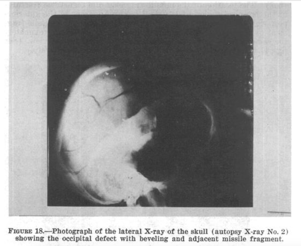
- The information (e.g., the sella turcica) used to orient the image as a "right" lateral X-ray in the "computer-assisted" version) are not actually visible in the base "un-enhanced" image upon which the "computer assisted" version is purportedly based.
- The original HSCA caption describes it as showing the "occipital defect"--which is medical-speak for "hole at the back of the head."
- Note the stuff at the bottom of the image--the spinal column and the two mastoid processes on either side of it, which are cropped out of the "computer assisted" image.
The HSCA documents never specify whether this is the "right" lateral image or the "left" lateral image.
I believe it is the left lateral, which X-ray technician Jerrol Custer said he was able to "just barely" get. Note that with the absence of facial features, it is impossible to determine from the X-ray itself whether it is "right" or "left," which is why an AP (Anterior-Posterior) view is also made. More on the missing "facial features" in a moment.
Here is the HSCA "un-enhanced" lateral X-ray, with its original caption:
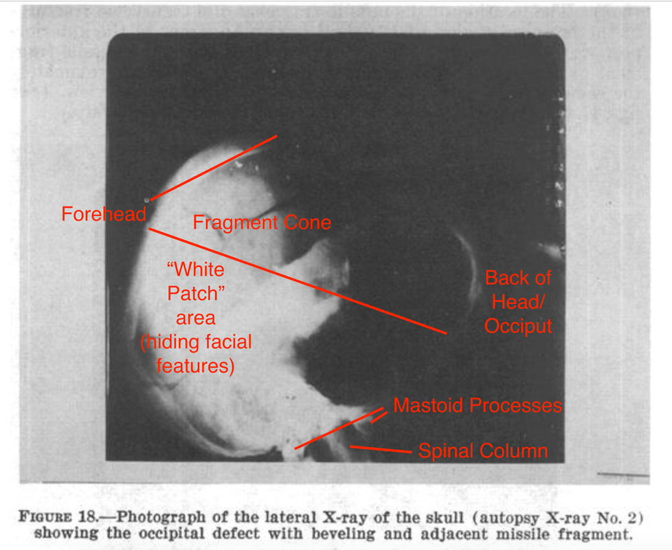
If you orient this X-ray so that the black area, the "defect" or "hole" is in the occipital region of the head as the caption describes, it would put the face towards the lower-left corner of the image--which is right where radiologist Dr. David Mantik describes the impossibly white "white patch." So while Dr. Chesser believed the dark area was "blackened to hide the facial features," I actually contend that the facial features were "whited out."
Here is my annotated version showing how this X-ray should be oriented (the lower part of the hole is where the Harper fragment, which ejected with the second head shot [AR-15 slam-fire shot] eventually came out.:
Here is my annotated version showing how this X-ray should be oriented (the lower part of the hole is where the Harper fragment, which ejected with the second head shot [AR-15 slam-fire shot] eventually came out.:
Note that neither Dr. Mantik nor Dr. Chesser was of the opinion that the X-ray images they viewed at NARA were "original" unaltered X-rays.
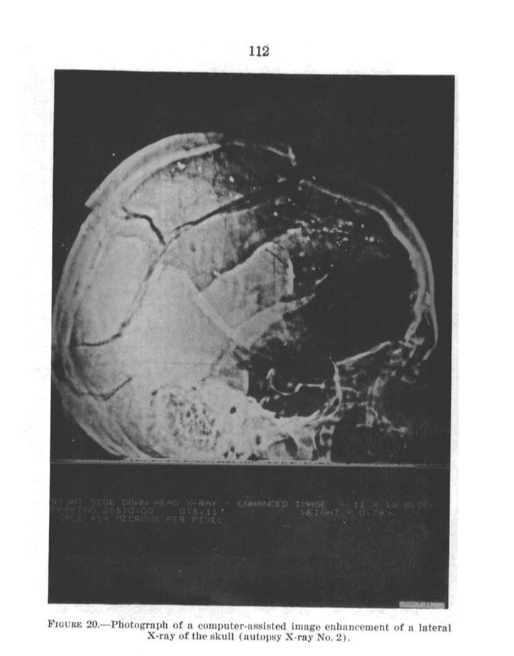
Note that two pages after the (un-enhanced) lateral X-ray image is presented in the HSCA documents, we find the so-called "computer assisted" or "computer enhanced" version of this lateral X-ray--the one everyone can easily find online--and now, all of the sudden, we have some features that were not visible in the original "un-enhanced" image. We also have an important part--specifically, the bottom of the "un-enhanced" original image--that is cropped off, so we no longer see the spinal column or the mastoid processes. One of the new things we can see, not visible in the original image, in in the middle of the dark area, where we now see a sickle-shaped feature, called the sella turcica. And on the right-most edge, we now see see some better-defined shapes that were not visible in the original.
Here is the "computer-assisted" version of "a lateral X-ray," supposedly a more highly defined version of the above X-ray:

The sella turcica and those more highly defined areas on the lower-right corner of the image now help us orient the face, because the lower-right corner area now looks like sinus cavities, and the sella turcica especially tells us which way front is--and it's not to the lower-left, as we would assume from the "occipital defect" caption in the original "un-enhanced" image, but now the face is on the lower-right.
Neurologist Dr. Michael Chesser has described the sella turcica on this image as being "too large." In fact, he describes the apparent bullet fragment trail as being "too high" for either the autopsy doctors' "EOP" entry location or the HSCA "cowlick" entry location. (more on that, in a moment.)
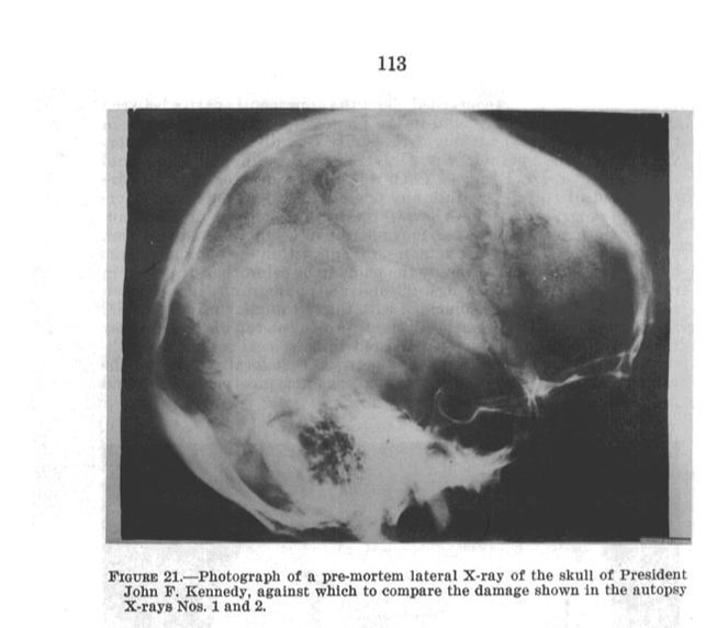
Interestingly, the HSCA also published, on the page immediately following this "computer-assisted" image, a "pre-mortem" lateral X-ray of Kennedy's head, taken when he was alive. And here's where things get even a bit more interesting...
If we compare the "pre-mortem" or "living" X-ray with the "computer-assisted" X-ray, we find a lot of similarities, landmarks that are exactly the same in both images, as if these X-rays were taken at exactly the same angle, from exactly the same distance, etc. In fact, I created a video overlaying the "computer-assisted" X-ray with the "living" X-ray to point out those similarities, and posted it on YouTube at https://www.youtube.com/watch?v=Q-CWAX__s10 where you can see how those landmarks line up exactly. The shape of the head, the sella turcica, and other landmarks align exactly.
However, when you overlay the "computer-assisted" lateral X-ray with the original "un-enhanced" lateral X-ray, as I did (see https://www.youtube.com/watch?v=92zCVZuJUWw for an overlay comparison of all three X-rays), much of that correspondence disappears. In fact, I had to do no stretching or re-sizing at all to get the "computer-assisted" image to align perfectly with the "living" X-ray. However, I did have to "stretch" the "un-enhanced" autopsy image in various dimensions to get the metallic fragments to align with the "computer-assisted" image, but no matter how I re-sized, stretched, adjusted, if I got one aspect of the un-enhanced X-ray to align with the same aspect in the "computer-assisted" X-ray, correspondence in other aspects was completely lost! And this is supposed to be the same X-ray!
You might be able to see what I'm talking about if you take a close look at the three X-rays side-by-side. Note the more "rounded" shape of the skull in the "un-enhanced" image on the right, than to the slightly more "squashed" shape of the skull in the "computer-assisted" image in the center, or the pre-mortem "living" X-ray on the left:
So what does all that mean? It means that the "computer-assisted" image is actually a composite image of the pre-mortem "living" X-ray on the left, and the "un-enhanced" autopsy image on the right.
Which is not to say that the "un-enhanced" autopsy image is entirely authentic. Dr. Mantik's "white patch" is still there. The "black hole" (which Dr. Chesser describes as having "blacked out" the facial features) is actually the occipital hole, but may have undergone a little dark room editing when the "white patch" was added. But instead of hiding the hole in the occiput, as Dr. Mantik theorized, the "white patch" is actually hiding the facial features (sinuses and orbits) rather than the hole at the back of the head described by all the Parkland and Bethesda witnesses.
Conclusions
The widely found "computer-assisted" or "computer-enhanced" lateral X-ray image, purporting to be the "right" lateral X-ray, is actually a composite of the "pre-mortem" or "living" X-ray, and the left lateral X-ray showing the "occipital defect" at the back of the head--right where all those Parkland and Bethesda witnesses describe the hole at the back of Kennedy's head. Dr. Mantik's "White Patch"--rather than hiding the occipital blow-out--actually hides the facial features (orbits and sinuses) that would otherwise have been visible and would have helped orient the X-ray correctly.
I posted the animation from my documentary, demonstrating how the un-enhanced left lateral X-ray actually shows the back-of-the-head blow-out on YouTube, "JFK Lateral Xray Animation" at https://www.youtube.com/watch?v=XKlbuP3uCuA. Please watch it, as it demonstrates visually what I am trying to explain in words.
And as for that bullet fragment trail that is "too high" to be from either an EOP or "cowlick" entrance--it's just the right height to be from the forehead entry location described by Dr. Charles Crenshaw, if one orients the front of the head at the lower left in the original "un-enhanced" autopsy image.


